Exploring the Benefits of Tape Stripping in Dermatology Clinical Research
| Table of contents |
| 1. RNA Analysis |
| 2. Protein Analysis |
| 3. Lipid Analysis |
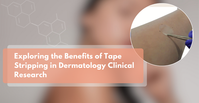
There are several approaches to study biomarkers using non-invasive techniques such as tape stripping and transdermal analysis patch. This article will focus on the tape stripping method.
The popularity of tape stripping in clinical trials has been steadily increasing for the last 19 years. This growing interest can be observed in the number of publications showcasing this non-invasive technique as well as the increasing number of clinical centers using this method to study the skin in drug and non-drug studies.
This method can be utilized to study RNA expression, gene expression, protein or lipids levels. Various techniques of tape stripping exist, these variations include different types of tapes, different application times, number of tapes, pressure, and extraction methods. These techniques all fall under the tape stripping method to suit the specific requirements of a study.

RNA Analysis
Case 1: atopic dermatitis and psoriasis study
This first case is from Innovaderm, featuring lab analysis by Dr. Guttman from the department of Dermatology at the Icahn School of Medicine at Mount Sinai in New York City, was recently published in the Journal of Allergy and Clinical Immunology where the focus was gene expression in patients with psoriasis and atopic dermatitis. Most available publications using tape strips were conducted in patients with atopic dermatitis; the objective of this specific study was to perform tape stripping in atopic dermatitis and psoriasis patients to study and compare the signals obtained.
This study was comprised of a total of 60 subjects: 20 subjects with atopic dermatitis, 20 subjects with psoriasis and 20 healthy subjects. Each subject had tape stripping with D-Squame (Cu-Derm). Twenty (20) tapes total were taken on normal-looking skin and on lesional skin of patients with atopic dermatitis and psoriasis and from skin of healthy subjects to extract RNA. The recovery rate for this procedure was very positive.
As previously demonstrated with skin biopsies, this publication shows the difference in gene expression between normal skin of atopic dermatitis patients and normal skin of non-atopic patients. The results also show a striking difference between normal skin and lesional skin of atopic dermatitis patients.
Th2: The study showed, just as it did for skin biopsies, that Th2 markers were increased in atopic dermatitis but not in psoriasis. Notably, IL-13 was increased in both normal skin and lesional skin of atopic dermatitis patients, IL-4 was increased in lesional skin, and IL-5 just as well.
Innate Immunity: Some increases were observed in innate immunity markers including iNOS that was only present in psoriasis skin. Since the increase was only seen in the skin of psoriasis patients, this could even be used as a diagnostic marker of psoriasis as compared to atopic dermatitis.
Th1: Th1 has some increases in psoriasis compared to atopic dermatitis. For example, the graph in the study presents Interferon gamma (IFN-γ) as higher for psoriasis than atopic dermatitis.
Th17: IL-17 is higher in psoriasis although it has some increase in lesional skin in atopic dermatitis as it has been reported before.
Some other markers like TARC have been reported to be elevated in both psoriasis and atopic dermatitis. IL-31 shows a strong signal in the itch cytokine in atopic dermatitis lesional skin which was not present in psoriasis.
Protein Analysis
Case 2: atopic dermatitis study
As previously mentioned, it is important to keep in mind that several tape stripping techniques exist. This following case is from a Japanese group that used a different variation of tapes called Cellotapes. This study required only 6 tapes to be collected. They measured IL-8 protein levels at different times after treatment with a topical corticosteroid showing a statistically significant decrease in IL-8 protein presence which paralleled clinical improvement.
Case 3: atopic dermatitis study
Dr. Clausen from Bispebjerg Hospital, Department of Dermatology , conducted a study utilizing D-Squame tapes to investigate protein extraction. The study focused on the protein content of samples when taken deeper into the epidermis comparing tapes from lesional skin and non-lesional skin of atopic dermatitis subjects and from healthy controls. It was shown that lesional skin, non-lesional skin and healthy control, could get more proteins per tape when they started at the stratum corneum; these numbers went down by the time they entered the lower levels of the epidermis.
However, when they looked at 3 chemokines, specifically TARC, CXCL8 and TSLP, the levels were fairly flat, suggesting that the levels of those chemokines can be measured even in the superficial layers of the epidermis.
Lipid Analysis
Case 4: atopic dermatitis study
Lipid analysis is a process that could be interesting in atopic dermatitis but has received more interest in acne vulgaris.
This last study by Dr. Leung’s group, from National Jewish Health, who has extensive experience and expertise in measuring lipids from the skin of patients with atopic dermatitis, was comprised of 30 adult patients with atopic dermatitis and 25 adult controls without atopic dermatitis.
They collected 20 consecutive tape strips and used only 2 strips for lipid analysis. Using those 2 strips, they demonstrated significant differences in certain ceramides between patients with atopic dermatitis and normal controls: or between lesional skin of patients with atopic dermatitis and non-lesional skin. There was a decrease in some ceramides in patients with atopic dermatitis which have been reported before with other techniques.
As it has been demonstrated through the multiple psoriasis and atopic dermatitis cases presented in this article, that non-invasive methods such as tape stripping can be an interesting path to study biomarkers, particularly using RNA, protein, and lipid analysis.
About the Author:
Julie’s experience exceeds 10 years in the domains of clinical pharmacology, medical writing, and scientific affairs. This extensive professional journey has been predominantly within the dynamic CRO environment. Julie assumes a pivotal role as medical writer within Innovaderm’s Scientific Affairs Department.
Innovaderm’s expertise in tape stripping and other non-invasive biomarkers has positioned the CRO as a leader in the field of dermatology. Our proficiency in the study of biomarkers is supported by rigorous and quality-oriented scientific expertise.
Newsletter
Newsletter subscription resources